Monday, May 29, 2017

MOHS surgery for Skin Cancer

MOHS surgery

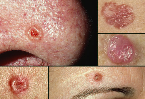
Developed by Dr. Frederick Mohs in the 1930s, Mohs micrographic surgery has come to be accepted as the single most effective technique for removing Basal Cell Carcinomas and Squamous Cell Carcinomas (BCCs and SCCs) (pic, 1) , the two most common skin cancers. It accomplishes the nifty trick of sparing the greatest amount of healthy tissue while also most completely expunging cancer cells; cure rates for BCC and SCC are an unparalleled 98 percent or higher with Mohs, significantly better than the rates for standard excision or any other accepted method.

The reason for the technique's success is its simple elegance. Mohs differs from other techniques in that microscopic examination of all excised tissues occurs during rather than after the surgery, thereby eliminating the need to "estimate" how far out or deep the roots of the skin cancer go. This allows the Mohs surgeon to remove all of the cancer cells while sparing as much normal tissue as possible. 

Basal.jpg

Picture 1: Basal Cell Carcinomas and Squamous Cell Carcinomas

In standard excisions, the visible skin cancer is removed along with a 4-8 mm margin of healthy appearing tissue (pic. 2).  The wound is then closed.  The tissue is then send out for the margins to be checked over the next few days.  When the pathologist looks at the tissue, they use a method called “bread loafing” where the pathologies makes slices through the tissue and examines those margins as a representative sample to determine that the margins are clear.

standard excision.jpg
Picture 2: Standard excision

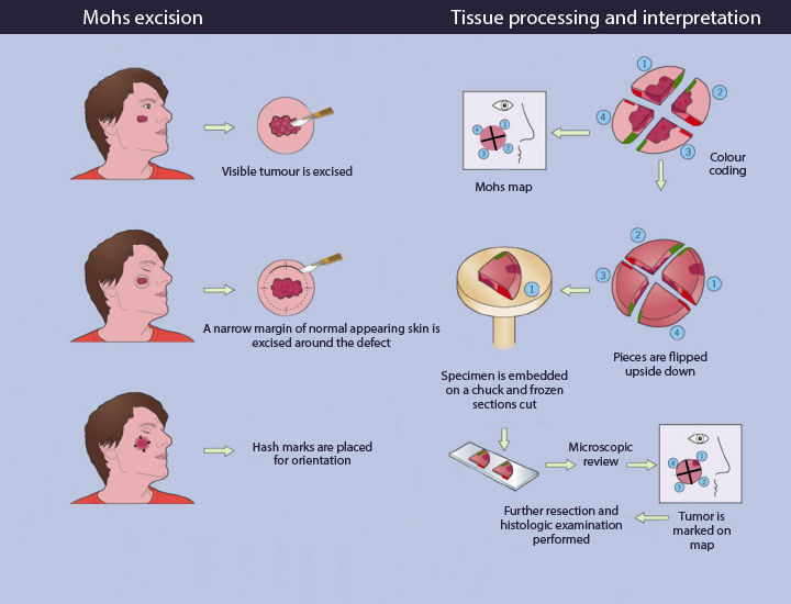
In Mohs surgeries, the visible cancer is removed along with a narrow rim of healthy tissue (Pic. 3). The tissue is then processed while the patient is waiting in the clinic. During the processing, the edges are laid down in order to examine 100% of the margins. If the margins are cancer-free, the surgery is ended. If not, more tissue is removed from the margin where the cancer cells were found, and the procedure is repeated until all the margins of the final tissue sample examined are clear of cancer. In this way, Mohs surgery eliminates the guesswork in skin cancer removal, producing the best therapeutic and cosmetic results.

MOHS.jpg

Picture 3: MOHS technique

Most common indications for MOHS surgery are:

 1) Primary BCC or SCC in an area which is important to preserve maximum healthy tissue for maximal functional and cosmetic result.  (e.g.  eyelids, Nose, ears, lips, genitals, hands, feet).

 2) Recurrent or incompletely excised BCC or SCC.

 3) Primary BCC or SCC where the margins cannot be clearly defined.

 4) Primary BCC or SCC larger than 2 cm or rapidly growing.

 5) Primary BCC or SCC arising in scars or in sites of radiation therapy.

 6) High risk or aggressive types of SCC.

 7) Patients with multiple tumors in the same surgical area.

 8) Young patients who may expect to have further skin cancers on the face in the future.
For more information

www.plasticsurgerycyprus.com
info@plasticsurgerycyprus.com
Tel: 95107711

Saturday, May 20, 2017

Χειρουργική Καρκίνου Δέρματος

Χειρουργική MOHS

Η μικρογραφική χειρουργική MOHS αναπτύχθηκε από τον Δρ Φρέντερικ Mohs το 1930 και έχει γίνει πλέον αποδεκτή, ως η πιο αποτελεσματική τεχνική για την αφαίρεση του Βασικοκυτταρικού καρκινώματος (BCC και Ακανθοκυτταρικού  καρκινώματος (εικ. 1) δέρματος. Με αυτή την τεχνική επιτυγχάνεται η ολοκληρωτική εξαίρεση της νεοπλασίας, εξοικονομώντας ταυτόχρονα υγειές ιστό. Οι έρευνες μιλούν για ποσοστό ίασης 98% ή και περισσότερο, σημαντικά υψηλότερο από άλλες τεχνικές αφαίρεσης.

Basal.jpg

εικ. 1: Βασικοκυτταρικά και Ακανθοκυτταρικά  καρκινώματα 

Η τεχνική Mohs διαφέρει από τις άλλες τεχνικές, στο γεγονός ότι η μικροσκοπική εξέταση όλων των ιστών γίνεται κατά την διάρκεια της επέμβασης και όχι μετά την χειρουργική επέμβαση. Έτσι  εξαλείφεται η ανάγκη να γίνει "εκτίμηση" αργότερα για το πόσο μακριά φθάνουν οι ρίζες του καρκίνου στο δέρμα. Έτσι διαφυλάσσετε όσο το δυνατό περισσότερος ιστός, που είναι πολύ σημαντικός για μια αισθητική και λειτουργική αποκατάσταση του ελλείματος.

standard excision.jpg
εικ. 2: Κλασσική εκτομή

Κατά την κλασσική εκτομή (εικ. 2) ,  ο ορατός καρκίνος του δέρματος αφαιρείτε μαζί με 4-6 mm υγειές ιστού ( ελλειπτική τομή). Γίνεται συρραφή του ελλείματος και το παρασκεύασμα αποστέλλετε για ιστολογική εξέταση τις προσεχείς ημέρες.  Ο ιστοπαθολόγος χρησιμοποιεί την τεχνική «Bread Loafing”. Η ιστολογική εξέταση γίνεται ουσιαστικά με κάθετες τομές σε αντιπροσωπευτικά διαστήματα μέσα στο δείγμα, για να καθοριστεί αν τα όρια είναι καθαρά.

MOHS.jpg

εικ. 3: Τεχνική MOHS 

Αντιθέτως στην τεχνική MOHS (εικ. 3)  ο ορατός καρκίνος του δέρματος αφαιρείτε μαζί με μικρό όριο υγειές ιστού. Ενώ ο ασθενής είναι σε αναμονή στην κλινική γίνετε μια ειδική επεξεργασία του ιστού. Ακολούθως γίνεται η σταδιακή εξέταση του παρασκευάσματος σε οριζόντιες τομές επιτρέποντας έτσι  την εξέταση του 100% των χειρουργικών ορίων σε τρεις διαστάσεις. Αν τα όρια είναι καθαρά τότε γίνετε σύγκλιση του ελλείματος. Αν όχι, τότε η διαδικασία επαναλαμβάνεται αφαιρώντας όμως μόνο την περιοχή που περιέχει καρκινικά κύτταρα, μέχρι να επιτευχθεί μία πλήρως αρνητική ιστολογική εξέταση.

Κυριότερες ενδείξεις για την τεχνική MOHS:

 1) Εντοπισμός του καρκίνου σε ζωτικής σημασίας ανατομικές μονάδες, όπου η εξοικονόμηση ιστού είναι σημαντική για το μέγιστο λειτουργικό  και αισθητικό αποτέλεσμα: περιoφθαλμικά, στην περιοχή της μύτης, περιστoματικά, στα αυτιά, στα γεννητικά όργανα και την περιoχή των χεριών και των ποδιών.

 2) Όγκοι με ασαφή κλινικά όρια ή εξελκωμένες βλάβες.

  3) Kαρκινώματα με διάμετρό μεγαλύτερη από 2cm.

 4) Yπoτρoπή του καρκίνου μετά τη θεραπεία ή αφαίρεση με θετικά όρια.

 5) Ακανθοκυτταρικά καρκινώματα υψηλού κίνδυνου η επιθετικής μορφής.

 6) Ασθενείς με πολλαπλούς όγκους στην ίδια χειρουργική περιοχή.

 7) Βασικοκυτταρικά και Ακανθοκυτταρικά  καρκινώματα που αναπτύσσονται σε ουλές ή σε περιοχές μετά από ακτινοθεραπεία.

 8) Σε νεαρούς ασθενείς αφού αναμένεται να έχουν περαιτέρω καρκίνους δέρματος στο πρόσωπο.


Για περισσότερες πληροφορίες:
www.plasticsurgerycyprus.com
info@plasticsurgerycyprus.com
Τηλ: 95107711