Monday, May 29, 2017

MOHS surgery for Skin Cancer

MOHS surgery

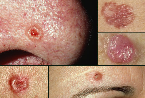
Developed by Dr. Frederick Mohs in the 1930s, Mohs micrographic surgery has come to be accepted as the single most effective technique for removing Basal Cell Carcinomas and Squamous Cell Carcinomas (BCCs and SCCs) (pic, 1) , the two most common skin cancers. It accomplishes the nifty trick of sparing the greatest amount of healthy tissue while also most completely expunging cancer cells; cure rates for BCC and SCC are an unparalleled 98 percent or higher with Mohs, significantly better than the rates for standard excision or any other accepted method.

The reason for the technique's success is its simple elegance. Mohs differs from other techniques in that microscopic examination of all excised tissues occurs during rather than after the surgery, thereby eliminating the need to "estimate" how far out or deep the roots of the skin cancer go. This allows the Mohs surgeon to remove all of the cancer cells while sparing as much normal tissue as possible. 

Basal.jpg

Picture 1: Basal Cell Carcinomas and Squamous Cell Carcinomas

In standard excisions, the visible skin cancer is removed along with a 4-8 mm margin of healthy appearing tissue (pic. 2).  The wound is then closed.  The tissue is then send out for the margins to be checked over the next few days.  When the pathologist looks at the tissue, they use a method called “bread loafing” where the pathologies makes slices through the tissue and examines those margins as a representative sample to determine that the margins are clear.

standard excision.jpg
Picture 2: Standard excision

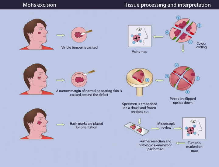
In Mohs surgeries, the visible cancer is removed along with a narrow rim of healthy tissue (Pic. 3). The tissue is then processed while the patient is waiting in the clinic. During the processing, the edges are laid down in order to examine 100% of the margins. If the margins are cancer-free, the surgery is ended. If not, more tissue is removed from the margin where the cancer cells were found, and the procedure is repeated until all the margins of the final tissue sample examined are clear of cancer. In this way, Mohs surgery eliminates the guesswork in skin cancer removal, producing the best therapeutic and cosmetic results.

MOHS.jpg

Picture 3: MOHS technique

Most common indications for MOHS surgery are:

 1) Primary BCC or SCC in an area which is important to preserve maximum healthy tissue for maximal functional and cosmetic result.  (e.g.  eyelids, Nose, ears, lips, genitals, hands, feet).

 2) Recurrent or incompletely excised BCC or SCC.

 3) Primary BCC or SCC where the margins cannot be clearly defined.

 4) Primary BCC or SCC larger than 2 cm or rapidly growing.

 5) Primary BCC or SCC arising in scars or in sites of radiation therapy.

 6) High risk or aggressive types of SCC.

 7) Patients with multiple tumors in the same surgical area.

 8) Young patients who may expect to have further skin cancers on the face in the future.

For more information

www.plasticsurgerycyprus.com
info@plasticsurgerycyprus.com
Tel: 95107711

Saturday, May 20, 2017

Χειρουργική Καρκίνου Δέρματος

Χειρουργική MOHS

Η μικρογραφική χειρουργική MOHS αναπτύχθηκε από τον Δρ Φρέντερικ Mohs το 1930 και έχει γίνει πλέον αποδεκτή, ως η πιο αποτελεσματική τεχνική για την αφαίρεση του Βασικοκυτταρικού καρκινώματος (BCC και Ακανθοκυτταρικού  καρκινώματος (εικ. 1) δέρματος. Με αυτή την τεχνική επιτυγχάνεται η ολοκληρωτική εξαίρεση της νεοπλασίας, εξοικονομώντας ταυτόχρονα υγειές ιστό. Οι έρευνες μιλούν για ποσοστό ίασης 98% ή και περισσότερο, σημαντικά υψηλότερο από άλλες τεχνικές αφαίρεσης.

Basal.jpg

εικ. 1: Βασικοκυτταρικά και Ακανθοκυτταρικά  καρκινώματα 

Η τεχνική Mohs διαφέρει από τις άλλες τεχνικές, στο γεγονός ότι η μικροσκοπική εξέταση όλων των ιστών γίνεται κατά την διάρκεια της επέμβασης και όχι μετά την χειρουργική επέμβαση. Έτσι  εξαλείφεται η ανάγκη να γίνει "εκτίμηση" αργότερα για το πόσο μακριά φθάνουν οι ρίζες του καρκίνου στο δέρμα. Έτσι διαφυλάσσετε όσο το δυνατό περισσότερος ιστός, που είναι πολύ σημαντικός για μια αισθητική και λειτουργική αποκατάσταση του ελλείματος.

standard excision.jpg
εικ. 2: Κλασσική εκτομή

Κατά την κλασσική εκτομή (εικ. 2) ,  ο ορατός καρκίνος του δέρματος αφαιρείτε μαζί με 4-6 mm υγειές ιστού ( ελλειπτική τομή). Γίνεται συρραφή του ελλείματος και το παρασκεύασμα αποστέλλετε για ιστολογική εξέταση τις προσεχείς ημέρες.  Ο ιστοπαθολόγος χρησιμοποιεί την τεχνική «Bread Loafing”. Η ιστολογική εξέταση γίνεται ουσιαστικά με κάθετες τομές σε αντιπροσωπευτικά διαστήματα μέσα στο δείγμα, για να καθοριστεί αν τα όρια είναι καθαρά.

MOHS.jpg

εικ. 3: Τεχνική MOHS 

Αντιθέτως στην τεχνική MOHS (εικ. 3)  ο ορατός καρκίνος του δέρματος αφαιρείτε μαζί με μικρό όριο υγειές ιστού. Ενώ ο ασθενής είναι σε αναμονή στην κλινική γίνετε μια ειδική επεξεργασία του ιστού. Ακολούθως γίνεται η σταδιακή εξέταση του παρασκευάσματος σε οριζόντιες τομές επιτρέποντας έτσι  την εξέταση του 100% των χειρουργικών ορίων σε τρεις διαστάσεις. Αν τα όρια είναι καθαρά τότε γίνετε σύγκλιση του ελλείματος. Αν όχι, τότε η διαδικασία επαναλαμβάνεται αφαιρώντας όμως μόνο την περιοχή που περιέχει καρκινικά κύτταρα, μέχρι να επιτευχθεί μία πλήρως αρνητική ιστολογική εξέταση.

Κυριότερες ενδείξεις για την τεχνική MOHS:

 1) Εντοπισμός του καρκίνου σε ζωτικής σημασίας ανατομικές μονάδες, όπου η εξοικονόμηση ιστού είναι σημαντική για το μέγιστο λειτουργικό  και αισθητικό αποτέλεσμα: περιoφθαλμικά, στην περιοχή της μύτης, περιστoματικά, στα αυτιά, στα γεννητικά όργανα και την περιoχή των χεριών και των ποδιών.

 2) Όγκοι με ασαφή κλινικά όρια ή εξελκωμένες βλάβες.

  3) Kαρκινώματα με διάμετρό μεγαλύτερη από 2cm.

 4) Yπoτρoπή του καρκίνου μετά τη θεραπεία ή αφαίρεση με θετικά όρια.

 5) Ακανθοκυτταρικά καρκινώματα υψηλού κίνδυνου η επιθετικής μορφής.

 6) Ασθενείς με πολλαπλούς όγκους στην ίδια χειρουργική περιοχή.

 7) Βασικοκυτταρικά και Ακανθοκυτταρικά  καρκινώματα που αναπτύσσονται σε ουλές ή σε περιοχές μετά από ακτινοθεραπεία.

 8) Σε νεαρούς ασθενείς αφού αναμένεται να έχουν περαιτέρω καρκίνους δέρματος στο πρόσωπο.


Για περισσότερες πληροφορίες:
www.plasticsurgerycyprus.com
info@plasticsurgerycyprus.com
Τηλ: 95107711

Thursday, March 24, 2016

Ear Plastic Surgery

Otoplasty
What are protruding ears?
Protruding ears are the most frequent disorders of the ears. Prominent ears are ears that are perceived as sticking out too far from the side of the head and can cause a severe psychological problem in men and women, boys and girls. A lot of people find different ways to cover them with their hair and they even avoid to go to the beach or expose their ears.
Otoplasty (plastic surgery of the ear) is the term used for the correction technique. People who are concerned about the appearance of their ears may eventually resort to otoplasty surgery to correct any perceived prominence.  It is considered to be a routine surgery performed by most of plastic surgeons.
What is the problem?
The problem of a protruding ear is mainly anatomical. Specifically, in a natural ear, helix and antihelix should be smooth and form an angle of 90 °. In protruding ears, this angle is not well-defined (greater than 90 °) and often the antihelix is absent, thus giving the image of a prominent ear. A second cause is the presence of large and deep concha, thereby pushing the ear out and forward. 
At what age should someone with protruding ears undergo surgery? 
The surgery in children, can be done in the immediate pre-school age of 5-6 years. This can help prevent any psychological trauma that teasing at school can cause. In adults, there is no age restriction.
What kind of anesthesia is used? 
The ear surgery can be done under local anesthesia. Local anesthetic is injected into the ear so it will get numb for about two to three hours. Very thin needles are used to reduce pain that may be caused during the injection. Thus, the intervention of otoplasty is a virtually painless procedure.
However, if the patient does not cooperate (usually children between 5-12 years old) sedation is required or even general anesthesia. Otoplasty can be performed as an outpatient. This means that on the day of plastic surgery you may leave the hospital or the clinic.
Which technique is used?
There are several techniques with many variations that can be used. Correction can be carried out only with sutures, with the removal of cartilage, thinning or scratching of cartilage or a combination of all the above.


You should be aware that the more invasive a technique is, the greater the risk for complications. It has not been established scientifically, the superiority of a technique over the other regarding long term results yet. For this reason, it is always attempted to correct the problem with sutures (the less invasive method) and only in special cases by removing or scratching the cartilage.
Price:
The total cost of the Otoplasty in Cyprus under local anesthesia in the office is between 1400-1500 euros.

For more Information


www.plasticsurgery4cyprus.com

info@plasticsurgerycyprus.com


Tel: 95107711

Friday, December 4, 2015

Breast Augmentation


What are Breast Implants?
A breast implant is a medical prosthesis made of medical silicon used to augment, reconstruct, or recreate the physical form of breasts.

Anatomical versus Round Implants?
Round implants are used more commonly as they feel softer and the orientation does not matter, making the surgery much easier. In very thin patients with little breast volume though, will make the breast look unnatural.

Anatomical implants look like teardrops and they can give the breast a natural shape. They can only be oriented in one way, so the pocket has to be very precise in order to avoid rotation leaving the breast looking misshaped. The implant will feel firmer as it is more solid in order to keep its shape.

Under or over the muscle?
With sub-glandular placement the implant location is between your chest muscle and your breast tissue (under the glands). This placement may help reduce your surgery and recovery time. It may be less painful than sub-muscular placement and may make your breast implants easier to access if reoperation is necessary. Implants that are sub-glandular may be easier to see and feel through your skin.

Sub-muscular placement means that your breast implants are placed partially or wholly under the pectoralis major chest muscle. Sub-muscular implants may result in a longer, more painful recovery than sub-glandular implants. Sub-muscular placement may result in less palpable implants and may reduce the risk of developing capsular contracture, as well as offer easier mammographic imaging of the breast.

For how long?
New breast implants do have the possibility of lasting a lifetime. They need to be changed only if you have problems with them such as capsular contracture, rupture or displacement. However due to aging, pregnancies and breast feeding, the breast tissue will lose its elasticity and sag, while the implant remains at the original location. That’s why a second operation might need to elevate the breast after several years.

Which size?
You should not choose the size based on the size of your best friends implants or your favorite actress. Each breast is unique and needs a unique evaluation. The breast implant is for the whole life and not for a couple of months. The bigger the size the more the complications the less will last. Your plastic surgeon will measure your breast and let you know the most appropriate size for you.

Any serious complications?
The most feared complication is the capsular contracture (hardening of the breast area around the implant). This can cause pain and deform the breasts.  Its frequency may range between 5-15% off all breast enlargements and in severe cases a second operation will be needed to change the implants. Other complications that they may need a second surgery are hematomas (collection of blood), implant displacement and infections. Rupture of the breast implant (a tear or hole in the implant) it’s a rare complication. There are no proven associations between implants and connective tissue diseases, autoimmune diseases, breast cancer or reproductive problems.


Overall breast enlargement is a safe procedure but you should aim for a small natural breast and should assume that you will need to have additional surgeries.

Price:
The cost of surgery can range between 3900-4200 euros.


For more Information

info@plasticsurgerycyprus.com
Tel: 95107711

Thursday, November 26, 2015

Γυναικομαστία

Γυναικομαστία


Τι είναι η γυναικομαστία;

Η γυναικομαστία είναι η ανάπτυξη στους άνδρες μεγάλου στήθους. Αυτό μπορεί να τους προκαλέσει προβλήματα σχετικά με την εμφάνισή τους, επηρεάζοντας παράλληλα και την ανδρική αυτοπεποίθησή τους. Πολλοί ασθενείς αποφεύγουν να εκθέτουν το στήθος τους στην παραλία, στο γυμναστήριο κ.λ.π. Επίσης, περιορίζει τις επιλογές τους στο ντύσιμο, αποφεύγοντας ρούχα που είναι πολύ εφαρμοστά. Ιδιαίτερα στην Κύπρο, μπορεί να προκαλέσει αρκετά ψυχολογικά προβλήματα κατά την διάρκεια της στρατιωτικής θητείας του έφηβου. 

gynecomastia.png

Αιτιολογία:

Η πιο συχνή μορφή είναι η λεγόμενη εφηβική γυναικομαστία. Εμφανίζεται σε ποσοστό 40% των εφήβων, σε ηλικίες μεταξύ 13 και 17 ετών και είναι αμφοτερόπλευρη (και τα δυο στήθη είναι μεγάλα). Στην πλειοψηφία όμως το πρόβλημα παρέρχεται μετά από 12-24 μήνες. Σε περιπτώσεις που η γυναικομαστία επιμένει, η χειρουργική θεραπεία είναι ίσως η μοναδική επιλογή.

Η λήψη αναβολικών επίσης μπορεί να οδηγήσει στην εμφάνιση της γυναικομαστίας και συνήθως αυτή παραμένει ακόμη και αν ο ασθενής σταματήσει την λήψη τους. Άλλες πιο σπάνιες αιτίες είναι η ηπατική και νεφρική ανεπάρκεια, λήψη φαρμάκων όπως διουρητικά η κατάχρηση αλκοόλ και ναρκωτικών. 

Ο καρκίνος του στήθους στους άντρες που είναι μια σπάνια πάθηση, μπορεί να εμφανιστεί με την μορφή της γυναικομαστίας. Συνήθως όμως εμφανίζετε μόνο στο ένα στήθος, μπορεί να προκαλέσει ανωμαλίες του δέρματος στην περιοχή που βρίσκεται και να συνοδεύεται από αιματηρές εκκρίσεις από τη θηλή.

Χρειάζεται κάποια Διερεύνηση;

Στις περιπτώσεις της εφηβικής γυναικομαστία, συνήθως δεν χρειάζεται κάποια ιδιαίτερη διερεύνηση. Ένας έμπειρος πλαστικός χειρουργός, μπορεί να καταλάβει αν πρόκειται για κάποιο παθολογικό η ορμονικό πρόβλημα. Σε τέτοιες περιπτώσεις θα χρειαστεί ένας ορμονικός έλεγχος. Στις σπάνιες περιπτώσεις, όπου υπάρχει η υποψία για καρκίνο θα χρειαστεί περαιτέρω ακτινολογική διερεύνηση. 

Ποιοι βαθμοί υπάρχουν;

Η ταξινόμηση της γυναικομαστίας είναι πολύ σημαντική για την επιλογή της θεραπείας. Υπάρχουν τέσσερεις βαθμοί:

  • Βαθμός I: Μικρός, ορατός διογκωμένος μαστός, χωρίς χαλάρωση του δέρματος.
  • Βαθμός IIA: Μέτρια διογκωμένος μαστός, χωρίς χαλάρωση του δέρματος.
  • Βαθμός IIB: Μέτρια διογκωμένος μαστός με χαλάρωση του δέρματος.
  • Βαθμός III: Αρκετά διογκωμένος μαστός, με μεγάλη χαλάρωση του δέρματος
gynecomastia cyprus.png

male breast enlargment cyprus.png

Ποια είναι η κατάλληλη θεραπεία; 

Η θεραπεία εξαρτάται από τον βαθμό του προβλήματος. Στους τύπους Ι, ΙΙΑ και μερικές φορές στον τύπο ΙΙΒ, η γυναικομαστία αντιμετωπίζεται με μια απλή λιποαναρρόφηση( η διαδικασία περιγράφεται στην επόμενη σελίδα). Με την λιποαναρρόφηση δεν αφαιρείτε μόνο το λίπος αλλά και μέρος του αδένα που προκαλεί το πρόβλημα. 

Σε πιο προχωρημένα στάδια όπου υπάρχει περίσσεια δέρματος χρειάζονται να γίνουν κάποιες τομές για την αφαίρεση του δέρματος. Αναλόγως του προβλήματος, οι τομές μπορεί να γίνουν  γύρω από την  θηλή, στην περιοχή του στήθους ή στην περιοχή της μασχάλης. 

Στους περισσότερους ασθενείς οι ουλές δεν είναι τόσο ορατές και δεν αποτελούν σοβαρό πρόβλημα. Δυστυχώς όμως σε κάποιες περιπτώσεις οι ουλές δεν σβήνουν εντελώς και μπορεί να είναι αρκετά ορατές. Για αυτό τον λόγο γίνετε προσπάθεια το πρόβλημα να αντιμετωπίζετε αρχικά μόνο με λιποαναρρόφηση που δεν αφήνει τομές. Ο ασθενής έχει έτσι την δυνατότητα να αποφασίσει σε αργότερο στάδιο αν θα προχωρήσει σε αφαίρεση του δέρματος ή όχι. Πολλές φόρες με την λιποαναρρόφηση επιτυγχάνετε τέτοια συρρίκνωση του δέρματος που το τελικό αποτέλεσμα είναι αρκετά ικανοποιητικό. 


Το ολικό κόστος της θεραπείας της Γυναικομαστίας με την μέθοδο της λιποαναρρόφησης ανέρχεται στα 2000 ευρώ.

Για περισσότερες πληροφορίες:

info@plasticsurgerycyprus.com


Τηλ: 95107711

Wednesday, November 18, 2015

Βλεφαροπλαστική άνω βλεφάρων

Η βλεφαροπλαστική είναι μια από τις συχνότερες επεμβάσεις που εκτελούνται στην πλαστική χειρουργική. Κατά την επέμβαση αφαιρείται υπερβολικό δέρμα καθώς επίσης και μερικές φορές λίπος από το άνω βλέφαρο.

Τα μάτια μας είναι από τα πρώτα σημεία στα οποία φαίνονται τα σημάδια του χρόνου, αφού καθώς περνάει η ηλικία εμφανίζεται περίσσεια δέρματος ή και πρόπτωση λίπους στα άνω βλέφαρα με αποτέλεσμα να φαινόμαστε κουρασμένοι.

Η επέμβαση γίνεται συνήθως με τοπική αναισθησία . Με την χρήση δύο λαβίδων καθορίζεται η περίσσειας του δέρματος που πρέπει να αφαιρεθεί. Η τομή γίνεται στις φυσικές πτυχές του βλεφάρου, έτσι ώστε η ουλή να μην είναι τόσο ορατή. Η περίσσεια του δέρματος αφαιρείται και αν είναι αναγκαίο το λίπος. Χρησιμοποιούνται πολύ λεπτά ράμματά για να μην μένουν σημάδια.


Συνήθως ο χρόνος αποκατάστασης είναι σύντομός και κάποιος ή κάποια μπορεί να κυκλοφορεί άμεσα φορώντας γυαλιά ηλίου. Μπορεί να υπάρξουν μώλωπες και πρήξιμο, αλλά εξαφανίζονται σε λίγες μέρες.  Η χρήση παγοκύστεων κατά τις πρώτες 48 ώρες μπορεί να επιταχύνει την επούλωση. Τα ράμματα αφαιρούνται μετά από 5-7 μέρες. Οι περισσότεροι ασθενείς επιστρέφουν στις κανονικές τους δραστηριότητες μετά από 7-10 ημέρες .


Μετά την επέμβαση, τα μάτια θα γίνουν πιο ξεκούραστα και θα αποκτήσουν μια ζωντάνια, αποκτώντας έτσι ένα καλύτερο σχήμα. 

Το ολικό κόστος της πλαστικής αυτιών με τοπική αναισθησία στο ιατρείο, ανέρχεται στα 1300 ευρώ.

Για περισσότερες πληροφορίες:

www.plasticsurgerycyprus.com
info@plasticsurgerycyprus.com
Τηλ: 95107711


Wednesday, November 11, 2015

Upper Lid Blepharoplasty


An eyelid lift, or blepharoplasty, is a surgical procedure to remove skin and fat from the eyelids. The term "eyelid lift" is a misconception because the eyelid is not lifted during surgery. 

Aging, gravity, loss of elastic tissue in the skin, and weakening of the connective tissues of the eyelid, frequently contribute to lax and redundant eyelid tissue. This excess tissues hangs down over the front edge of the eyelidWeakening of the tissue can also cause protrusion of some eye fat presenting as puffiness in the medial upper eyelid.

These changes will result in a heavy looking eyelid, aged appearance, puffiness, and sometimes will impair the vision.  

Upper eyelid blepharoplasty is performed under local anesthesia. By pinching the excess skin between two forceps, the amount of vertical skin excision is determined. The excess skin is excised and the protruding fat is removed. The incision lines are made in the natural creases of the upper lids so any resulting scarring is hidden. The wound is closed with fine sutures so the scars will become less noticeable and troublesome.


Usually patients have a short recovery time after blepharoplasty. Although bruising and swelling is usually worse on the day after surgery, they quickly begin to disappear. During the first 48 hours the use of cool compresses can greatly reduce bruising and swelling around the eyes and face. Light activity such as walking will also help speed healing. The stitches can remain in the skin for about a week. Most people return to normal activities after 7 to 10 days following blepharoplasty. By 2 weeks after eyelid surgery, the majority of the bruising and swelling will resolve. 

A successful eyelid surgery can make your eyes look younger and more alert while providing you with enhanced self-esteem and confidence in your appearance. 

Price: 
The cost for upper lid Blepharoplasty under local anesthesia is 1300 euros. 

For more information: 

info@plasticsurgerycyprus.com 
Τηλ: 95107711